The Aging Pupil: What's Quietly Happening Inside Your Eyes Long Before You Notice
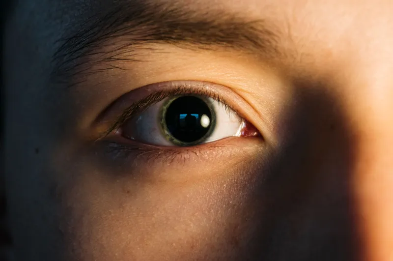
When most Americans think about age-related vision changes, they picture reading glasses on a nightstand or squinting at a computer screen. Rarely does the conversation turn to something far more fundamental — the pupil itself. Yet the small, dark circle at the center of your eye undergoes a slow and largely invisible transformation with age, one that quietly shapes how well you see in nearly every lighting condition you encounter throughout your day.
This is not a minor cosmetic shift. The pupil's behavior — how wide it opens in darkness, how tightly it contracts in bright light, and how quickly it transitions between the two — governs the quality and comfort of your vision in ways that standard prescriptions cannot fully address.
What the Pupil Actually Does — and Why It Matters More Than Most People Realize
The pupil is the eye's aperture, functioning much like the opening in a camera lens. In dim environments, it dilates to allow more light to reach the retina. In bright conditions, it constricts to protect the retina from overexposure and to sharpen focus. This dynamic range is controlled by two sets of muscles in the iris: the dilator muscle, which widens the pupil, and the sphincter muscle, which narrows it.
In a healthy young adult, the pupil can expand to roughly 7 or 8 millimeters in complete darkness. By the time a person reaches their sixties, that maximum dilation often shrinks to somewhere between 4 and 5 millimeters — a reduction that may sound modest but translates into a significant decrease in the amount of light entering the eye. Less incoming light means reduced contrast sensitivity, diminished depth perception in low-light settings, and a greater struggle to adapt when moving from a bright space into a darker one.
The term clinicians sometimes use for this age-related reduction in maximum pupil size is senile miosis, though the condition is neither rare nor dramatic in its onset. It develops gradually over decades, which is precisely why so many people fail to connect their worsening night vision to this specific mechanism.
The Neurological Story Behind Pupil Aging
Pupil behavior is not solely a mechanical phenomenon. It is neurologically governed, with the autonomic nervous system — specifically the sympathetic and parasympathetic branches — directing the iris muscles in real time. As the nervous system ages, signal transmission slows, and the speed with which the pupil can respond to changing light conditions decreases alongside it.
This lag has real-world consequences. Consider the experience of driving at night when an oncoming vehicle's headlights suddenly flood your visual field. A younger pupil constricts rapidly to limit the glare and then re-dilates quickly once the car passes, restoring night vision within seconds. An older pupil responds more sluggishly in both directions — it is slower to constrict against the glare and slower to recover afterward. That recovery window, which may extend to fifteen seconds or more in older adults compared to just a few seconds in younger ones, is precisely when the road ahead becomes most difficult to see.
Beyond the speed of response, the neural pathways responsible for pupillary light reflexes can also be affected by systemic conditions common in older Americans — including diabetes, hypertension, and certain neurological disorders — making the picture considerably more complex for patients managing multiple health concerns.
Why Standard Eye Exams May Not Catch This Early Enough
A routine eye examination typically assesses visual acuity, refractive error, intraocular pressure, and the health of the retina and optic nerve. While a clinician will often check the pupillary light reflex as part of a neurological screening, the nuanced measurement of maximum pupil dilation under various lighting conditions and the speed of the pupillary response across different light transitions is not a standard component of most annual exams.
This means that a patient can walk out of an appointment with a perfectly updated prescription and still experience significant difficulty driving after dusk — a discrepancy that can be frustrating and, in some cases, genuinely dangerous. Patients who raise these concerns are sometimes told their prescription is correct and their retinas appear healthy, which is accurate but incomplete. The problem, in those cases, may lie not in what the eye sees but in how much light it is able to gather in the first place.
If you find yourself struggling with night driving, experiencing halos or starbursts around headlights, or needing considerably more light than you once did to read comfortably, it is worth asking your eye care provider specifically about pupillary function and whether a more comprehensive low-light assessment might be appropriate.
Practical Adjustments That Can Make a Meaningful Difference
While the aging of the pupil cannot be reversed, several evidence-informed strategies can help manage its effects and preserve visual comfort across different environments.
Prioritize anti-reflective lens coatings. For those who wear prescription eyeglasses, high-quality anti-reflective coatings reduce the amount of light lost to reflection on the lens surface — a meaningful benefit when the pupil is already limiting how much light reaches the retina. This is particularly valuable for nighttime driving.
Allow adequate time for dark adaptation. Because older pupils adapt more slowly, building in deliberate transition time when moving from bright to dim environments can reduce the disorientation and visual uncertainty that often accompanies these shifts. Before stepping out of a brightly lit building into a dark parking lot, a brief pause near the exit can allow the eye to begin its adjustment.
Optimize indoor lighting strategically. Rather than relying on a single overhead fixture, layered lighting — combining ambient, task, and accent sources — can reduce the dramatic contrast between bright and dark areas within a room. This lessens the demand placed on the pupil to make frequent, rapid adjustments.
Discuss medication interactions with your physician. A number of commonly prescribed medications, including certain antihistamines, antidepressants, and blood pressure drugs, can affect pupil size and response speed. If you have noticed changes in your light sensitivity or night vision after starting a new medication, that conversation is worth having.
Protect your eyes from excessive UV exposure. Chronic sun exposure is associated with accelerated changes in the lens and other anterior eye structures, which can compound the effects of pupillary aging. Consistent use of UV-blocking sunglasses outdoors remains one of the most straightforward protective measures available.
Reframing the Conversation Around Vision and Aging
There is a tendency in American health culture to treat vision changes as either correctable with a new prescription or simply inevitable and therefore not worth examining closely. The truth, as with most aspects of health, is more nuanced. Understanding the specific mechanisms through which the eye ages — including the often-overlooked decline in pupillary function — allows patients to make more informed decisions about their care, ask more precise questions during appointments, and adopt habits that genuinely support long-term visual performance.
The pupil may be small, but its role in shaping the quality of your vision is anything but minor. Paying attention to how your eyes behave in different lighting conditions, and communicating those observations clearly to your eye care provider, is one of the most meaningful steps you can take toward maintaining the clarity and safety your vision deserves.