Floaters, Flashes, and the Signals Your Eyes May Be Sending You
It happens without warning. You glance toward a bright window or a pale stretch of sky, and there it is — a small, drifting shape at the edge of your vision. It moves when your eyes move, retreats when you try to focus on it, and disappears as quickly as it arrived. Or perhaps you notice a brief, brilliant flash of light in your peripheral field, despite the room being dimly lit. These experiences are among the most common visual phenomena reported to eye care professionals, and they generate a considerable amount of anxiety among patients who encounter them for the first time.
Understanding what floaters and flashes are, why they occur, and — critically — when they require immediate medical attention is knowledge that every adult should carry.
What Are Eye Floaters, Exactly?
The human eye is filled with a gel-like substance called the vitreous humor, which occupies the large interior chamber between the lens and the retina. In youth, this gel is uniformly clear and firmly attached to the retinal surface. As the eye ages — typically beginning in the fourth or fifth decade of life — the vitreous gradually liquefies and its internal protein fibers begin to clump together. These aggregated fibers cast shadows on the retina, and those shadows are what the brain perceives as floaters.
The forms floaters take can vary considerably. Some appear as small dots or specks. Others manifest as thin, thread-like strands, irregular cobweb shapes, or rings. The characteristic feature of all floaters is their tendency to drift across the visual field when the eye moves and then continue to shift slightly even after the eye has stopped — a result of the floater's inertia within the fluid vitreous.
Floaters are more visible against uniform, bright backgrounds — a cloudless sky, a white wall, or a well-lit computer screen — and less perceptible in low-contrast or dimly lit environments.
The Mechanics Behind Flashes of Light
Flashes — brief, lightning-like streaks or arcs of light perceived in the peripheral visual field — arise through a different mechanism, though they are closely related to the same underlying process that produces floaters.
The retina, which lines the interior back surface of the eye, contains photoreceptor cells that respond to light stimulation. However, those cells can also be activated by mechanical stimulation — specifically, by being tugged or pulled. When the vitreous gel shrinks and pulls against the retina, it generates a mechanical stimulus that the brain interprets as light, even in complete darkness. This is why flashes are often described as occurring in the periphery and are particularly noticeable in dim conditions or when the eyes are closed.
Flashes tend to be brief — lasting a fraction of a second — and may recur intermittently over a period of days or weeks during periods of active vitreous movement.
Posterior Vitreous Detachment: The Most Common Cause
The most frequent explanation for a sudden onset of floaters and flashes in adults, particularly those over the age of 50, is a condition called posterior vitreous detachment, or PVD. This occurs when the vitreous gel separates from the retinal surface, which it had previously been adhered to throughout the patient's life. The process is a natural part of aging and is estimated to affect the majority of people by the age of 70.
In most cases, PVD is entirely benign. The floaters it produces may be noticeable and initially bothersome, but they typically become less intrusive over weeks to months as the brain learns to filter them out. The flashes associated with PVD generally subside once the vitreous has fully separated and is no longer tugging on the retinal surface.
However, PVD is not always uncomplicated. In a minority of cases — estimates from ophthalmological literature suggest roughly 10 to 15 percent — the vitreous does not release cleanly and instead pulls with sufficient force to tear the retina.
When Floaters and Flashes Become a Medical Emergency
A retinal tear is a serious development. If fluid from within the vitreous cavity passes through the tear and accumulates beneath the retina, the retina can begin to separate from the underlying tissue — a condition known as retinal detachment. Retinal detachment is a medical emergency. Without prompt surgical intervention, it can lead to permanent, irreversible vision loss.
The warning signs that distinguish a potentially dangerous situation from a benign one include:
- A sudden, dramatic increase in the number of floaters, particularly if dozens or hundreds of new spots appear at once
- Flashes of light that are persistent, frequent, or increasing in intensity
- A shadow, curtain, or dark veil that appears to encroach on any portion of the visual field
- A sudden blurring or distortion of central vision
Any of these symptoms warrants same-day evaluation by an eye care professional. This is not a situation in which a patient should adopt a wait-and-see approach or schedule a routine appointment for the following week. Emergency rooms can assess acute vision changes, but an optometrist or ophthalmologist with dilated fundus examination capability is the appropriate destination when retinal pathology is suspected.
Individuals who are at elevated risk for retinal complications — including those who are highly myopic (nearsighted), have a personal or family history of retinal detachment, have undergone prior eye surgery, or have experienced significant eye trauma — should be particularly vigilant about reporting new visual symptoms promptly.
Other Conditions That Can Produce Similar Symptoms
Not all flashes of light originate in the eye itself. Ocular migraines, for instance, produce a distinctive visual aura that typically manifests as a shimmering, arc-shaped pattern of light — often described as a zigzag or fortification pattern — that expands slowly across the visual field over a period of 20 to 30 minutes. Unlike retinal flashes, ocular migraine aura affects both eyes simultaneously and is generally not associated with floaters. While sometimes alarming, ocular migraines are typically harmless from a neurological standpoint.
Flashes and visual disturbances can also arise from neurological causes, including transient ischemic attacks. If visual symptoms are accompanied by other neurological signs — such as numbness, weakness, difficulty speaking, or loss of balance — emergency evaluation is warranted.
What to Expect at Your Eye Appointment
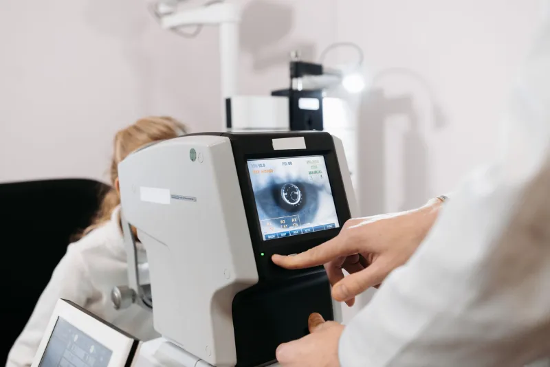
When a patient presents with new floaters or flashes, the standard of care involves a dilated fundus examination — a procedure in which the pupil is widened with eye drops to allow the clinician an unobstructed view of the vitreous, retina, and optic nerve. This examination is the only reliable method for ruling out retinal tears or detachment. It is painless, though patients should expect temporary light sensitivity and blurred near vision lasting several hours following dilation.
If a retinal tear is identified, it can often be treated in-office using laser photocoagulation or cryotherapy to seal the tear before detachment occurs. When detected early, these interventions carry a high success rate.
Living With Benign Floaters
For the many patients whose floaters are confirmed to be benign, the practical reality is that most will persist to some degree indefinitely. The vast majority of people find that their awareness of floaters diminishes significantly over time, as the brain effectively learns to disregard them. In rare cases where floaters are severely disruptive to vision and quality of life, surgical options such as vitrectomy exist, though they carry procedural risks that must be weighed carefully with an ophthalmologist.
The most important takeaway is this: do not dismiss sudden changes in your vision as something to simply tolerate. Your eyes are communicating information. The role of professional evaluation is to interpret that information accurately — and, when necessary, to act on it before a manageable situation becomes irreversible.